
Hip Fractures
Parrish Healthcare’s Hip Fracture Program
Our expert team provides personalized hip fracture care from surgery through rehabilitation, focusing on pain management, mobility and preventing future falls. With coordinated treatment, advanced techniques and compassionate support, we are proud to help patients return to independence and an improved quality of life as quickly as possible.
Understanding Hip Fractures
 The hip is a ball and joint socket. The socket is part of the pelvis and
is known as the acetabulum. The ball is the top part of the thigh bone
(femur) and is called the femoral head. The femoral head is attached to
the femur by the femoral neck. The greater trochanter is the bump that
you feel on the outside part of your hip. The lesser trochanter is a bump
on your femur bone that is lower than the greater trochanter and cannot
be felt by your hand. The trochanters are where many large muscles attach
to your hip and buttocks. Most often, an impact injury (such as falling)
is the cause of hip fractures. Hip fractures may also occur because of
osteoporosis. A hip fracture is a break in the top section of the femur
(thigh) bone.
The hip is a ball and joint socket. The socket is part of the pelvis and
is known as the acetabulum. The ball is the top part of the thigh bone
(femur) and is called the femoral head. The femoral head is attached to
the femur by the femoral neck. The greater trochanter is the bump that
you feel on the outside part of your hip. The lesser trochanter is a bump
on your femur bone that is lower than the greater trochanter and cannot
be felt by your hand. The trochanters are where many large muscles attach
to your hip and buttocks. Most often, an impact injury (such as falling)
is the cause of hip fractures. Hip fractures may also occur because of
osteoporosis. A hip fracture is a break in the top section of the femur
(thigh) bone.
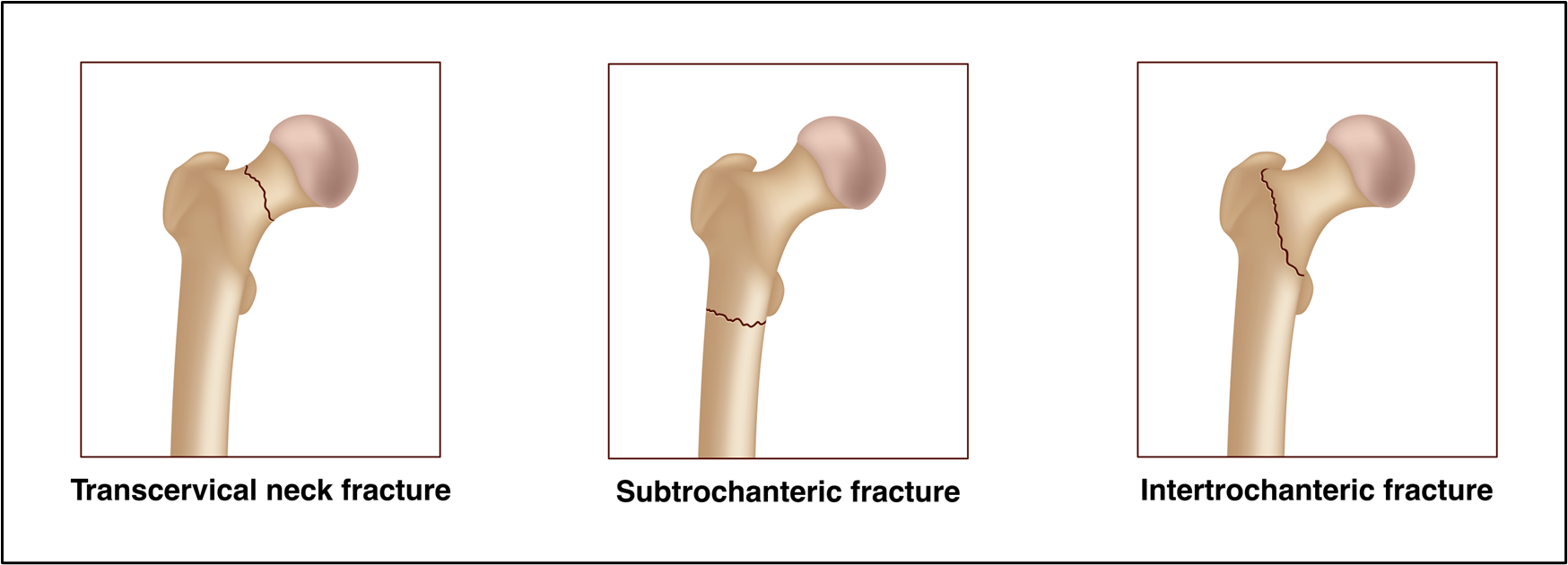
Types of Hip Fractures
There are a few ways your hip can break:
- Transcervical Fracture (Femoral Neck Fracture): In this type of fracture, the ball/femoral head breaks off the top of the femur.
- Intertrochanteric Fracture: This type of fracture is a break of the femur between the greater trochanter and the lesser trochanter.
- Subtrochanteric Fracture: In this type of fracture, the break in the femur occurs just below the trochanters, towards the top of the femur
Suspect a Hip Fracture?
If you or a loved one has experienced a fall and may have fractured a hip, please call 911 or immediately visit the nearest Emergency Department. If you or a loved one is already at the hospital, we encourage you to inquire with your care team about Parrish Healthcare’s Hip Fracture Program and how our team can support through surgery and recovery.
Diagnosing a Hip Fracture
An x-ray is used to diagnose hip fractures. X-rays will be used to determine if surgery is necessary and which type of surgical procedure is required to fix the fracture. Sometimes, an x-ray does not adequately show the fracture. In these cases, the doctor may order a Computer Tomography (CT) scan or a Magnetic Resonance Imaging (MRI) to get a better view of the bones and fracture pattern.
Treatment Options
Your orthopedic surgeon can begin to treat your hip fracture once you are medically stable. At that time, we will discuss with you the type of fracture that has occurred and your treatment options. Several surgical repair processes include:
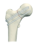
- Metal Screws (Refer to Diagram A below): In a femoral neck fracture where pieces of the bone are still in alignment, metal screws are used to hold the fractured pieces of the bone together.
- Metal Plate and Screws: For intertrochanteric fractures or subtrochanteric fractures, specifically designed plates and screws are used to hold together the broken fragments of bone.
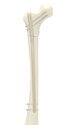
- Intramedullary Nail (Refer to Diagram B below)/Gamma Nail (Refer to Diagram C below): For intertrochanteric or subtrochanteric fractures, a metal rod is placed inside the bone, and screws are used to connect the fragments of the fracture.
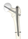
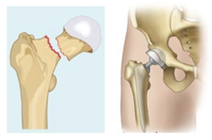
- Partial Hip Replacement (Hemiarthroplasty) or Full Hip Replacement (Total Hip Arthroplasty) (Refer to Diagram D below): In a femoral neck fracture where the pieces of the bone are no longer aligned, there is a high risk of interruption of blood supply to the head of the femur. When this occurs, the fracture cannot be fixed. The surgeon will remove the existing femoral head and replace it with a metal ball.
The decision for a total hip verse a hemi arthroplasty is dependent on the patient’s age and activity level. The total recovery for hip fracture surgery is typically 6-8 weeks (about 2 months).
Prior to surgery, our team will meet with you to discuss hospital care and what to expect the day of surgery and following surgery.
|
|
|
|
|
|
Diagram A Metal Screws |
Diagram B Intramedullary Nail |
Diagram C Gamma Nail |
Diagram D Bipolar Hip |
Post Hip Fracture Surgery Care
Following treatment of a hip fracture, we will work with you on both physical and occupational therapy goals. All therapy opportunities are designed to support your healing journey. Depending on the severity of your fracture and the quality of your bone, your surgeon will also determine your weight-bearing status. That is, whether you are allowed to put all your weight on your operated leg (full weight-bearing) or part of your weight on your operated leg (partial weight-bearing). Either way, you will also be advised of a walking aid to help support your hip. This includes the proper use of a walker, using a walker on curbs and/or stairs, getting in and out of bed, using a chair and toilet, dressing, bathing, getting in and out of a car, driving and traveling.
Home exercises are also essential for a complete recovery from hip fracture surgery. Some exercises focus on improving your range of motion and flexibility while other exercises focus on restoring you to your full strength after surgery. Your therapist, whether at home or at a facility, will help customize a plan for your individual needs. For optimal results, we encourage our patients to participate in an ongoing home exercise program in addition to the therapy provided by a therapist.
To learn more about Parrish Healthcare’s Hip Fracture Program and how we can support you or a loved one through surgery and recovery, please call our offices at (321) 268-6868.